2. 北京市缓控释肥料工程技术研究中心 北京 100097
2. Research Center of Beijing Municipal Slow and Controlled Release Fertilizers Engineering Technology, Beijing 100097, China
废弃塑料的普遍存在及诸多负面影响使公众感到不安, 这让塑料污染成为了环境领域研究的焦点问题。2004年, 英国科研人员在《科学》上发表的一篇论文中首次提出微塑料概念[1], 微塑料污染开始进入大众视线。目前尚未有全球公认的微塑料定义。塑料按尺寸大小可分为:大(macroplastic, > 2.5 cm)、中(mesoplastic, 5 mm~2.5 cm)、微(microplastic, MP 100 nm~5 mm)和纳(nanoplastic, < 100 nm)[2-3]。微塑料具体又可分为:小(small, < 1 mm)、中(medium, 1~3 mm)和大(large, 3~5 mm)[4]。近期, 一些学者建议窄化微塑料的范围(1~1000 µm), 将尺寸小于1 µm的塑料归为纳米塑料[5-6]。微塑料的形貌类型包括微珠(beads)、颗粒(nurdles或pellets)、纤维(fibers)、泡沫(foams)、碎片(fragments)和膜(films)等[7-8]。大部分微塑料的化学结构稳定、难降解、可长期存在(或以更小尺寸的形式存在), 将对生态环境安全和人类健康带来潜在的威胁。形貌特征直接影响微塑料在土壤中的迁移行为[9-10]。环境风险与尺寸、组分和含量等因素息息相关, 微塑料尺寸越小, 环境风险越大, 对其的检测越困难。受采样、前处理和分析技术的限制, 现有研究中检测到的微塑料尺寸普遍较大, 其实际数目被明显低估[11-12]。另外, 文献数据之间的可比性差, 不容易达成共识。沉积物中微塑料的分析方法可以经过微调后用于土壤微塑料的分析。不过, 对于土壤微塑料而言, 复杂的组分和表面附着物的存在导致分析检测存在更大的挑战, 尚难以形成统一、规范的标准方法[13]。
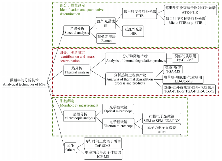
微塑料的成分本质上是聚合物, 对其的研究检测主要借助于分析聚合物的技术从组分、形貌(包括颜色、形状和尺寸)、含量(包括数量和质量)等几个维度进行, 如图 1所示。本文对这些分析技术在土壤微塑料研究中的应用现状进行解析、对比及总结, 以期对今后土壤微塑料分析技术的完善、标准化以及相关研究的开展有所借鉴。

|
图 1 微塑料的分析技术 Fig. 1 Analytical techniques of microplastics (MPs) |
通过将微塑料样品的光谱图与光谱库中的已知光谱进行比对获得具体的结构信息, 从而确定微塑料的化学组分, 并统计得出微塑料的数量, 是一种非破坏性分析方法。使用该方法检测微塑料样品时, 通常将其与显微镜技术结合、成像技术与化学计量学方法结合, 从而实现对样品的自动、快速定性定量分析。
1.1 红外光谱法(Infrared Spectroscopy, IR)红外光谱是吸收光谱, 信息从分子对入射电磁波的吸收得到。目前常见的红外光谱法有傅里叶变换红外光谱法(FTIR)和近红外光谱法(NIR)。
FTIR操作简单, 谱图的特征性强, 既可以用于环境中微塑料的原位识别和定量[14-15], 又可以用于老化微塑料的分析[16-17]。FTIR可以自由切换反射、透射、衰减全反射(ATR)模式, 满足对不同样品的分析需求。ATR-FTIR可直接快速测试肉眼可见的尺寸不小于200 µm的微塑料[18-20]。为了保证图谱的效果, 通常检测的微塑料尺寸不小于1 mm。傅里叶变换显微红外光谱法(micro-FTIR或者µ-FTIR)是将显微镜装在傅里叶变换红外光谱仪上, 既是微量分析又是微区分析的一种方法。该方法可以满足对粒径大于10 µm[21](也有文献提到粒径限值为20 µm[22])的微塑料的检测要求。不过, 检测过程中每次只能分析土壤样本中挑拣出的1个微塑料颗粒, 这样操作过程繁琐。配备焦平面(FPA)阵列或线阵列检测器后, µ-FTIR可实现对整个滤膜上的微塑料的全自动测量, 成为了目前用于免预分类、可视化、节省时间、自动识别、定量测定微塑料的最常用方法[23-24], 这为微塑料检测方法标准的建立奠定了基础。缺点是该方法需要去除目标物以外的大部分物质。为此, 样品的前处理尤为重要[25]。另外, 考虑到该方法检测到的微塑料数量会被低估[26], 与其他分析技术联用可以探索更多的化学信息, 这样检测精准度提高、范围扩大。比如:原子力显微镜红外光谱联用(AFM-IR)有望对尺寸更小的微塑料, 甚至纳米塑料进行表征[16, 27]。
NIR是利用有机基团在近红外区的电磁波的吸收特性来快速检测样品[28], 已广泛应用于塑料回收[29]、材料检测[30]及药物分析[31]等领域, 在微塑料研究中的应用刚刚起步, 尤其是土壤微塑料方面的报道有限[32]。Piarulli等[33]采用近红外高光谱图像技术(NIR-HIS)对水中3种标准聚合物进行了结构分析, 发现该法可以免去手动预挑选, 直接识别滤膜上尺寸小至80 µm的微塑料颗粒, 从而大大减少操作和分析时间以及潜在破坏或丢失颗粒的风险。Corradini等[34]采用可见光-近红外光谱法(vis-NIR)对模拟的土壤微塑料含量进行预测时发现, 该方法免去了提取过程, 直接量化样品中微塑料的含量, 可以满足快速检测土壤微塑料的需求, 适用于微塑料污染区。Paul等[35]采用高通量快速无损近红外光谱法对模拟的土壤微塑料进行检测时也证实该方法可用于较大样本量的在线预测。Qiu等[36]进一步将迁移学习法和NIR结合用于快速分析土壤微塑料, 建立了能同时评价不同区域土壤微塑料污染水平的分级模型。发现在分析微塑料时迁移学习方法特别是MEDA算法比传统的多变量分析方法更有效。
1.2 拉曼光谱法(Raman spectroscopy)除了红外光谱法, 拉曼光谱法是另外一种在微塑料的原位组分定性和颗粒统计中发挥重要作用的方法[37-38]。拉曼光谱是散射光谱, 信息从入射光与散射光频率的差别得到。与红外光谱法相比, 拉曼光谱法具有更高的空间分辨率, 不受水分干扰等优点, 一些红外光谱不能检测的信息可通过拉曼光谱获得。缺点在于如果样品产生荧光效应或者添加剂比较多时会影响样品检测, 或者检测不到拉曼信号。另外, 当微塑料尺寸比较小时, 测试样品有被激光损坏的风险。当与显微镜联用时, 显微拉曼光谱(micro-Raman或者µ-Raman)可以对10 µm以下、1 µm以上的微塑料进行表征[39]。拉曼成像技术是将共聚焦显微镜、拉曼光谱技术和新型信号探测装置相结合, 能够快速获得高信噪比的未经有机质消解的微塑料的拉曼图像[40]。Frère等[41]提出了一种结合静态图像分析的半自动显微拉曼光谱分析方法。该方法能快速检测环境微塑料的形貌及化学结构信息, 颗粒识别率在70%以上。另外, 可以在样品区域上采用面扫模式自动逐点采集信号, 大大提高检测效率和准确率[42-43]。有文献认为, 未去除的有机干扰物质对分析结果准确率的影响是红外光谱法和拉曼光谱法的共性[44]。将拉曼和红外结合使用, 可提高微塑料的鉴定准确率[45]。
2 热分析热分析是另一种研究微塑料的常用方法, 是针对微塑料热降解产物进行测定的破坏性分析方法。根据降解产物结构的片段信息推断微塑料组分和结构, 并给出质量分数。与光谱法相比, 该方法受粒径限制小, 受干扰物质少。不足之处在于无法提供数量和形貌信息。而且, 由于该方法的聚合物数据库仍在开发中, 不如光谱法完备, 因此精准的定量分析仍需时间[46]。Fojt等[47]提出该方法在检测土壤中的生物降解微塑料(micro-bioplastics)时最有前景。该方法具体分为两种:其一, 仅对热降解产物进行分析, 裂解气质联用法(Py-GC-MS); 其二, 对热解过程和产物均进行跟踪分析, 热重波谱联用法, 如热重-气质联用(TGA-GC-MS)、萃取-热脱附-气质联用(TED-GC-MS)和热重-红外(TGA-FTIR)。
2.1 裂解气质联用Py-GC-MS是在惰性气体环境中, 采用热裂解器将微塑料裂解, 受热裂解的产物通过色谱分离, 进入质谱仪离解, 得到粒子碎片, 从而确定聚合物结构[48-51], 是一种灵敏且成熟的适用于多数聚合物及其有机物的定性定量方法。其显著优点是同时识别聚合物和有机添加剂如塑化剂, 有助于将微塑料的产生环境与其来源联系起来, 并提供有关其年龄和衍变的有用信息[52]。其缺点在于, 热裂解仓的尺寸和允许上机样品量非常小, 分别为1.5 mm和0.5 mg, 对异质或成分复杂样品如土壤微塑料的重复性差, 所以在准备样品时要格外小心。其次, 因为不同的微塑料可能产生相似的热裂解产物, 存在误判风险。为了降低定量限, Dierkes等[53]将加压液体萃取(PLE)和Py-GC-MS结合开发了一种可用于土壤微塑料的定量分析方法。萃取包括甲醇预萃取、四氢呋喃萃取两步。样品中聚乙烯(PE)和聚丙烯(PP)的检测含量范围为0.03~3.3 mg∙g−1。该方法具有灵敏度高, 检测速度快, 通用性强等特点, 通过自动化有望实现批量样品分析。
2.2 热重波谱联用热重研究的是样品质量随温度变化的规律。土壤中的微塑料和其他组分不同(有时官能团类似), 失重曲线也不同。根据热解温度可以准确预测微塑料的一致性。另外, 一般情况下聚合物在500 ℃左右基本全部分解[54], 对于600~800 ℃之间的失重, 可以判断为微塑料表面附着物或填料的分解。因此, 热重波谱联用能快速确定微塑料、附着物或填料的类型和含量。
具体的热重波谱联用技术包括:热重-质谱(TGA-MS)[55]、热萃取-热脱附-气质联用(TED-GC- MS)、热重-红外(TGA-FTIR)和TGA-FTIR-GC-MS。热重-固相萃取(TGA-solid-phase extraction)与热脱附-气质(TDS-GC-MS)合称为TED-GC-MS, 即将热重分析和微塑料降解固相萃取产物的气相色谱、质谱分析相结合[56]。Dümichen等[57]报道称这种方法允许上机样品量可达100 mg, 保证了样品的均匀性, 能够测量复杂组分样品(如土壤微塑料), 并能够快速对其进行定性定量, 测试时间为2~3 h。文献中对于在测试时是否需要预处理的说法不统一[40]。同样, TGA-FTIR也可以增加样品的辨识度。Yu等[58]考察了TGA-FTIR检测环境微塑料的可行性, 采用该方法对土壤微塑料聚氯乙烯(PVC)和聚苯乙烯(PS)进行了定量分析。张玉佩等[59]制定了聚酰胺(PA)标准样品的TGA-FTIR定量标准曲线, 用实际海水样品验证了该方法的有效性。不过, 在分析微塑料样品时发现, 有微塑料热解物质红外信号相互重叠的现象存在, 这增加了微塑料定量分析的难度。余建平[60]基于TGA-FTIR-GC-MS联机技术定量分析了环境中常见的PE、PP、PVC等5种微塑料, 并指出对于不同环境样品, 需要选择适当的指示剂及前处理方法, 否则会造成检测结果偏差。热重-差示扫描量热法(TGA-DSC)也可提供微塑料PE和PP粒子碎片的信息[61], 并实现微塑料的定量分析。
3 显微技术显微技术是观察和研究微塑料形貌的最直接的方法。具体应用的有两种:一种是目视法中常用到的光学显微镜, 另一种是分辨率更高的电子显微镜。
3.1 光学显微镜光学显微镜可以提供微塑料的形貌信息, 在肉眼挑选微塑料的过程中起着举足轻重的作用, 即采用人工计数的方法计算出微塑料颗粒数目, 再换算成样品中的含量。优点在于操作简单、测试成本低。检测尺寸极限为500 μm。随着微塑料尺寸减小、透明度增加, 以及干扰物存在, 肉眼辨别微塑料的误判率增加[62-63]。例如, Song等[64]指出, 采用光学显微镜分析时, 碎片状微塑料显著低估, 纤维状微塑料显著高估。因此, 该方法不能作为检测微塑料的单独方法。
3.2 电子显微镜电子显微镜是显微技术的一个重要分支, 其核心是显示肉眼所不能直接看到的微塑料。它是利用电子束对样品放大成像的一种显微镜。目前微塑料研究中用到的主要有扫描电子显微镜(scanning electron microscopy, SEM)和原子力显微镜(atomic force microscopy, AFM)两种。
SEM是用二次电子加背景散射电子成像。放大倍数高, 成像清晰, 分辨率可达0.1 μm。颗粒表面的高分辨率图像可帮助研究者从有机颗粒中识别出微塑料[65]。扫描电子显微镜-能量色散X射线联用(SEM-EDS/EDX)可以同时对微塑料进行表面形貌鉴定和元素组成分析, 因此可以从无机粒子中辨别出微塑料。Gniadek等[66]比较了SEM-EDS/EDX与别的分析方法的优缺点, 认为用SEM-EDS/EDX分析海水微塑料时更有效、简单, 更重要的是它有望在纳米塑料的表征中发挥更大的作用。Wang等[67]提到了其他分析方法无法比拟的优点, 即SEM图像可显示出微塑料表面的裂纹和色素。SEM-EDS/EDX可直接给出微塑料表面覆盖着生物膜、放射虫和甲壳类动物等成分的证据, 可以识别出矿物质及含独特元素的微塑料(如氯化塑料)。不过, SEM要求样品是导电且平整的, 这使其应用受到很大限制。AFM的设计类似于SEM技术, 它们的许多主件相同。不同的是, AFM可以在任何环境(如液体、空气)中成像, 且超越了光和电子波长对显微镜分辨率的限制, 可在三维立体上在纳米级、分子级水平上观察样品的形貌和尺寸, 即该方法有望实现纳米塑料的形貌表征[16, 27]。
4 其他目前应用于环境微塑料的分析方法越来越丰富。飞行时间二次离子质谱(ToF-SIMS)作为一种独特的质谱技术, 适用于分析无机元素和有机化合物, 甚至复杂分子, 可进行快速质谱扫描和特征有机离子成像。它能够提供颗粒尺寸及分布信息。Du等[68-69]利用ToF-SIMS快速鉴定出土壤中PP、PVC、聚对苯二甲酸乙二醇酯(PET)和聚(酰胺6, PA6)4种微塑料的存在, 并获得了粒径分布和数量丰度信息。河北保定市12个典型区域土壤中PET和PA6占比较大, 达30%以上, 粒径分布在35 μm以下。电感耦合等离子体质谱(ICP-MS)是另一种高效质谱分析方法。Bolea-Fernandez等[70]尝试用单粒子ICP-MS检测粒径为1 μm和2.5 μm的聚苯乙烯(PS)微球, 成功获得了环境中两种模型微塑料的尺寸分布和质量分数等信息。Elert等[71]在比较红外、拉曼、TED-GC-MS和液相色谱(分子排阻色谱SEC)4种分析方法时提到SEC是一种快速定量评估环境中微塑料的有效方法。X射线衍射技术也是一种有价值的表征手段。Turner[72]使用便携式X射线荧光光谱仪(XRF)分析了环境微塑料中的元素组成, 所含微塑料的尺寸和含量可低至亚微米和亚毫克级。不过这些方法目前大部分仅限于模拟或其他环境体系, 还未用于真实土壤微塑料检测中。
5 总结与展望从光谱分析、热分析和显微分析等角度对土壤微塑料研究中应用的检测方法进行了综述。光谱分析对微塑料进行定性并统计数量, 常见的有傅里叶变换红外光谱法和拉曼光谱法。热分析用于组分和质量分析, 具体分为裂解气质联用和热重波谱联用。显微分析则对形貌和尺寸进行表征, 包括光学显微镜和电子显微镜两种。虽然应用于土壤微塑料的分析技术越来越丰富, 但是针对土壤微塑料的鉴别分析是一项复杂的工作, 仍存在一些问题需要解决:
1) 分析技术尚无统一的标准, 这样会大大降低相关研究之间的可比性, 分析技术标准的缺乏成为了制约该领域发展的关键因素。因此, 迫切需要制定包括土壤微塑料定性识别、定量分析在内的统一规程, 从而提高检测分析数据的可靠性与可比性。
2) 现有的土壤微塑料检测方法各有利弊。单独的检测方法无法准确获得微塑料的信息, 分析技术的组合或联用有望高效、准确地实现对土壤微塑料定性定量分析。除了对微塑料本身进行研究外, 分析技术的组合或联用还有助于获得微塑料中的添加剂、微塑料与其他污染物的耦合作用等信息, 更有助于清晰了解微塑料的来源。
3) 在分析前应仔细考虑需要解决的科学问题, 决定研究目的, 比如是对样品中微塑料颗粒的精准定性定量研究还是对区域土壤微塑料污染进行快速评估, 根据研究目的选择合适的分析技术。
4) 虽然土壤微塑料分析技术在不断改进或修正, 但是部分技术仅仅在室内模拟试验能获得一定的数据, 由于实验室模拟的环境与实际环境相差较大, 故针对实际土壤微塑料的测定, 仍有待于进一步的探索与验证。
| [1] |
THOMPSON R C, OLSEN Y, MITCHELL R P, et al. Lost at sea:where is all the plastic?[J]. Science, 2004, 304(5672): 838. DOI:10.1126/science.1094559 |
| [2] |
BLETTLER M C M, ULLA M A, RABUFFETTI A P, et al. Plastic pollution in freshwater ecosystems:macro-, meso-, and microplastic debris in a floodplain lake[J]. Environmental Monitoring and Assessment, 2017, 189(11): 581. DOI:10.1007/s10661-017-6305-8 |
| [3] |
HORTON A A, WALTON A, SPURGEON D J, et al. Microplastics in freshwater and terrestrial environments:evaluating the current understanding to identify the knowledge gaps and future research priorities[J]. Science of the Total Environment, 2017, 586: 127-141. DOI:10.1016/j.scitotenv.2017.01.190 |
| [4] |
QI R M, JONES D L, LI Z, et al. Behavior of microplastics and plastic film residues in the soil environment:a critical review[J]. Science of the Total Environment, 2020, 703: 134722. DOI:10.1016/j.scitotenv.2019.134722 |
| [5] |
GIGAULT J, HALLE A T, BAUDRIMONT M, et al. Current opinion:what is a nanoplastic?[J]. Environmental Pollution, 2018, 235: 1030-1034. DOI:10.1016/j.envpol.2018.01.024 |
| [6] |
HARTMANN N B, HÜFFER T, THOMPSON R C, et al. Are we speaking the same language? recommendations for a definition and categorization framework for plastic debris[J]. Environmental Science & Technology, 2019, 53(3): 1039-1047. |
| [7] |
HIDALGO-RUZ V, GUTOW L, THOMPSON R C, et al. Microplastics in the marine environment:a review of the methods used for identification and quantification[J]. Environmental Science Technology, 2012, 46(6): 3060-3075. DOI:10.1021/es2031505 |
| [8] |
WU P F, HUANG J S, ZHENG Y L, et al. Environmental occurrences, fate, and impacts of microplastics[J]. Ecotoxicology and Environmental Safety, 2019, 184: 109612. DOI:10.1016/j.ecoenv.2019.109612 |
| [9] |
DE SOUZA MACHADO A A, KLOAS W, ZARFL C, et al. Microplastics as an emerging threat to terrestrial ecosystems[J]. Global Change Biology, 2018, 24(4): 1405-1416. DOI:10.1111/gcb.14020 |
| [10] |
杨婧婧, 徐笠, 陆安祥, 等. 环境中微(纳米)塑料的来源及毒理学研究进展[J]. 环境化学, 2018, 37(3): 383-396. YANG J J, XU L, LU A X, et al. Research progress on the sources and toxicology of micro(nano)plastics in environment[J]. Environmental Chemistry, 2018, 37(3): 383-396. |
| [11] |
ZHANG B, YANG X, CHEN L, et al. Microplastics in soils:a review of possible sources, analytical methods and ecological impacts[J]. Journal of Chemical Technology & Biotechnology, 2020, 95(8): 2052-2068. |
| [12] |
GONG J, XIE P. Research progress in sources, analytical methods, eco-environmental effects, and control measures of microplastics[J]. Chemosphere, 2020, 254: 126790. DOI:10.1016/j.chemosphere.2020.126790 |
| [13] |
HURLEY R R, NIZZETTO L. Fate and occurrence of micro(nano)plastics in soils:knowledge gaps and possible risks[J]. Current Opinion in Environmental Science & Health, 2018, 1: 6-11. |
| [14] |
LI J, SONG Y, CAI Y B. Focus topics on microplastics in soil:analytical methods, occurrence, transport, and ecological risks[J]. Environmental Pollution, 2020, 257: 113570. DOI:10.1016/j.envpol.2019.113570 |
| [15] |
KUMAR M, XIONG X N, HE M J, et al. Microplastics as pollutants in agricultural soils[J]. Environmental Pollution, 2020, 265: 114980. DOI:10.1016/j.envpol.2020.114980 |
| [16] |
LUO H W, XIANG Y H, ZHAO Y Y, et al. Nanoscale infrared, thermal and mechanical properties of aged microplastics revealed by an atomic force microscopy coupled with infrared spectroscopy (AFM-IR) technique[J]. Science of the Total Environment, 2020, 744: 140944. DOI:10.1016/j.scitotenv.2020.140944 |
| [17] |
DING L, MAO R F, MA S R, et al. High temperature depended on the ageing mechanism of microplastics under different environmental conditions and its effect on the distribution of organic pollutants[J]. Water Research, 2020, 174: 115634. DOI:10.1016/j.watres.2020.115634 |
| [18] |
MATSUGUMA Y, TAKADA H, KUMATA H, et al. Microplastics in sediment cores from Asia and Africa as indicators of temporal trends in plastic pollution[J]. Archives of Environmental Contamination and Toxicology, 2017, 73(2): 230-239. DOI:10.1007/s00244-017-0414-9 |
| [19] |
FULLER S, GAUTAM A. A procedure for measuring microplastics using pressurized fluid extraction[J]. Environmental Science & Technology, 2016, 50(11): 5774-5780. |
| [20] |
SCOPETANI C, CHELAZZI D, MIKOLA J, et al. Olive oil-based method for the extraction, quantification and identification of microplastics in soil and compost samples[J]. Science of the Total Environment, 2020, 733: 139338. DOI:10.1016/j.scitotenv.2020.139338 |
| [21] |
WANG W F, GE J, YU X Y, et al. Environmental fate and impacts of microplastics in soil ecosystems:progress and perspective[J]. Science of the Total Environment, 2020, 708: 134841. DOI:10.1016/j.scitotenv.2019.134841 |
| [22] |
KÄPPLER A, FISCHER D, OBERBECKMANN S, et al. Analysis of environmental microplastics by vibrational microspectroscopy:FTIR, Raman or both?[J]. Analytical and Bioanalytical Chemistry, 2016, 408(29): 8377-8391. DOI:10.1007/s00216-016-9956-3 |
| [23] |
TAGG A S, SAPP M, HARRISON J P, et al. Identification and quantification of microplastics in wastewater using focal plane array-based reflectance micro-FT-IR imaging[J]. Analytical Chemistry, 2015, 87(12): 6032-6040. DOI:10.1021/acs.analchem.5b00495 |
| [24] |
LÖDER M G J, KUCZERA M, MINTENIG S, et al. Focal plane array detector-based micro-Fourier-transform infrared imaging for the analysis of microplastics in environmental samples[J]. Environmental Chemistry, 2015, 12(5): 563. DOI:10.1071/EN14205 |
| [25] |
SARKER A, DEEPO D M, NANDI R, et al. A review of microplastics pollution in the soil and terrestrial ecosystems:a global and Bangladesh perspective[J]. Science of the Total Environment, 2020, 733: 139296. DOI:10.1016/j.scitotenv.2020.139296 |
| [26] |
ZHANG Q, XU E G, LI J N, et al. A review of microplastics in table salt, drinking water, and air:direct human exposure[J]. Environmental Science & Technology, 2020, 54(7): 3740-3751. |
| [27] |
CHEN Y Y, WEN D S, PEI J C, et al. Identification and quantification of microplastics using Fourier-transform infrared spectroscopy:current status and future prospects[J]. Current Opinion in Environmental Science & Health, 2020, 18: 14-19. |
| [28] |
杨忠, 刘亚娜, 吕斌, 等. 非接触式可见光-近红外光谱法快速预测天然高分子材料表面粗糙度的研究[J]. 光谱学与光谱分析, 2013, 33(3): 682-685. YANG Z, LIU Y N, LYU B, et al. Rapid prediction of surface roughness of natural polymer material by visible/near infrared spectroscopy as a non-contact measurement method[J]. Spectroscopy and Spectral Analysis, 2013, 33(3): 682-685. DOI:10.3964/j.issn.1000-0593(2013)03-0682-04 |
| [29] |
ZHENG Y, BAI J R, XU J N, et al. A discrimination model in waste plastics sorting using NIR hyperspectral imaging system[J]. Waste Management, 2018, 72: 87-98. DOI:10.1016/j.wasman.2017.10.015 |
| [30] |
马翔.化学计量学结合近红外光谱在天然橡胶聚合物检测分析中的应用[D].北京: 北京化工大学, 2019 MA X. Application of chemometrics combined with NIR detection and analysis of NR polymers[D]. Beijing: Beijing University of Chemical Technology, 2019 |
| [31] |
白文明, 王来兵, 成日青, 等. 近红外高光谱成像技术在药物分析中的研究进展[J]. 药物分析杂志, 2018, 38(10): 1661-1667. BAI W M, WANG L B, CHENG R Q, et al. Research advance in pharmaceutical analysis based on near-infrared hyperspectral imaging technique[J]. Chinese Journal of Pharmaceutical Analysis, 2018, 38(10): 1661-1667. |
| [32] |
MÖLLER J N, LÖDER M G J, LAFORSCH C. Finding microplastics in soils:a review of analytical methods[J]. Environmental Science & Technology, 2020, 54(4): 2078-2090. |
| [33] |
PIARULLI S, SCIUTTO G, OLIVERI P, et al. Rapid and direct detection of small microplastics in aquatic samples by a new near infrared hyperspectral imaging (NIR-HSI) method[J]. Chemosphere, 2020, 260: 127655. DOI:10.1016/j.chemosphere.2020.127655 |
| [34] |
CORRADINI F, BARTHOLOMEUS H, LWANGA E H, et al. Predicting soil microplastic concentration using vis-NIR spectroscopy[J]. Science of the Total Environment, 2019, 650: 922-932. DOI:10.1016/j.scitotenv.2018.09.101 |
| [35] |
PAUL A, WANDER L, BECKER R, et al. High-throughput NIR spectroscopic (NIRS) detection of microplastics in soil[J]. Environmental Science and Pollution Research, 2019, 26(8): 7364-7374. DOI:10.1007/s11356-018-2180-2 |
| [36] |
QIU Z J, ZHAO S T, FENG X P, et al. Transfer learning method for plastic pollution evaluation in soil using NIR sensor[J]. Science of the Total Environment, 2020, 740: 140118. DOI:10.1016/j.scitotenv.2020.140118 |
| [37] |
SCHYMANSKI D, GOLDBECK C, HUMPF H U, et al. Analysis of microplastics in water by micro-Raman spectroscopy:release of plastic particles from different packaging into mineral water[J]. Water Research, 2018, 129: 154-162. DOI:10.1016/j.watres.2017.11.011 |
| [38] |
SARAU G, KLING L, OßMANN B E, et al. Correlative microscopy and spectroscopy workflow for microplastics[J]. Applied Spectroscopy, 2020, 74(9): 1155-1160. DOI:10.1177/0003702820916250 |
| [39] |
PRIMPKE S, CHRISTIANSEN S H, COWGER W, et al. Critical assessment of analytical methods for the harmonized and cost-efficient analysis of microplastics[J]. Applied Spectroscopy, 2020, 74(9): 1012-1047. DOI:10.1177/0003702820921465 |
| [40] |
刘婧.共聚焦显微拉曼光谱技术在海洋沉积物微塑料检测中的探索应用[D].北京: 中国科学院大学, 2020: 9-14 LIU J. Exploration and application of confocal micro-Raman spectroscopy in detection of marine sediment microplastics[D]. Beijing: University of Chinese Academy of Sciences, 2020: 9-14 |
| [41] |
FRÈRE L, PAUL-PONT I, MOREAU J, et al. A semi-automated Raman micro-spectroscopy method for morphological and chemical characterizations of microplastic litter[J]. Marine Pollution Bulletin, 2016, 113(1/2): 461-468. |
| [42] |
刘丹童, 宋洋, 李菲菲, 等. 基于显微拉曼面扫的小尺寸微塑料检测方法[J]. 中国环境科学, 2020, 40(10): 4429-4438. LIU D T, SONG Y, LI F F, et al. A detection method of small-sized microplastics based on micro-Raman mapping[J]. China Environmental Science, 2020, 40(10): 4429-4438. DOI:10.3969/j.issn.1000-6923.2020.10.029 |
| [43] |
ARAUJO C F, NOLASCO M M, RIBEIRO A M P, et al. Identification of microplastics using Raman spectroscopy:latest developments and future prospects[J]. Water Research, 2018, 142: 426-440. DOI:10.1016/j.watres.2018.05.060 |
| [44] |
BLÄSING M, AMELUNG W. Plastics in soil:analytical methods and possible sources[J]. Science of the Total Environment, 2018, 612: 422-435. DOI:10.1016/j.scitotenv.2017.08.086 |
| [45] |
BRANDT J, BRANDT J, BITTRICH L, et al. High-throughput analyses of microplastic samples using Fourier transform infrared and Raman spectrometry[J]. Applied Spectroscopy, 2020, 74(9): 1185-1197. DOI:10.1177/0003702820932926 |
| [46] |
HUPPERTSBERG S, KNEPPER T P. Instrumental analysis of microplastics-benefits and challenges[J]. Analytical and Bioanalytical Chemistry, 2018, 410(25): 6343-6352. DOI:10.1007/s00216-018-1210-8 |
| [47] |
FOJT J, DAVID J, PŘIKRYL R, et al. A critical review of the overlooked challenge of determining micro-bioplastics in soil[J]. Science of the Total Environment, 2020, 745: 140975. DOI:10.1016/j.scitotenv.2020.140975 |
| [48] |
FRIES E, DEKIFF J H, WILLMEYER J, et al. Identification of polymer types and additives in marine microplastic particles using pyrolysis-GC/MS and scanning electron microscopy[J]. Environmental Science Processes & Impacts, 2013, 15(10): 1949-1956. |
| [49] |
STEINMETZ Z, KINTZI A, MUÑOZ K, et al. A simple method for the selective quantification of polyethylene, polypropylene, and polystyrene plastic debris in soil by pyrolysis-gas chromatography/mass spectrometry[J]. Journal of Analytical and Applied Pyrolysis, 2020, 147: 104803. DOI:10.1016/j.jaap.2020.104803 |
| [50] |
KÄPPLER A, FISCHER M, SCHOLZ-BÖTTCHER B M, et al. Comparison of μ-ATR-FTIR spectroscopy and py-GCMS as identification tools for microplastic particles and fibers isolated from river sediments[J]. Analytical and Bioanalytical Chemistry, 2018, 410(21): 5313-5327. DOI:10.1007/s00216-018-1185-5 |
| [51] |
HERMABESSIERE L, HIMBER C, BORICAUD B, et al. Optimization, performance, and application of a pyrolysis-GC/MS method for the identification of microplastics[J]. Analytical and Bioanalytical Chemistry, 2018, 410(25): 6663-6676. DOI:10.1007/s00216-018-1279-0 |
| [52] |
GOMIERO A, ØYSÆD K B, AGUSTSSON T, et al. First record of characterization, concentration and distribution of microplastics in coastal sediments of an urban fjord in south west Norway using a thermal degradation method[J]. Chemosphere, 2019, 227: 705-714. DOI:10.1016/j.chemosphere.2019.04.096 |
| [53] |
DIERKES G, LAUSCHKE T, BECHER S, et al. Quantification of microplastics in environmental samples via pressurized liquid extraction and pyrolysis-gas chromatography[J]. Analytical and Bioanalytical Chemistry, 2019, 411(26): 6959-6968. DOI:10.1007/s00216-019-02066-9 |
| [54] |
张红, 刘鸿. 热分析技术在高分子材料研究中的应用[J]. 广州化工, 2001, 29(4): 39-42. ZHANG H, LIU H. Applications of thermal analysis technique on studies of polymer materials[J]. Guangzhou Chemical Industry, 2001, 29(4): 39-42. DOI:10.3969/j.issn.1001-9677.2001.04.014 |
| [55] |
DAVID J, STEINMETZ Z, KUČERÍK J, et al. Quantitative analysis of poly(ethylene terephthalate) microplastics in soil via thermogravimetry-mass spectrometry[J]. Analytical Chemistry, 2018, 90(15): 8793-8799. DOI:10.1021/acs.analchem.8b00355 |
| [56] |
DUEMICHEN E, BRAUN U, SENZ R, et al. Assessment of a new method for the analysis of decomposition gases of polymers by a combining thermogravimetric solid-phase extraction and thermal desorption gas chromatography mass spectrometry[J]. Journal of Chromatography A, 2014, 1354: 117-128. DOI:10.1016/j.chroma.2014.05.057 |
| [57] |
DÜMICHEN E, EISENTRAUT P, BANNICK C G, et al. Fast identification of microplastics in complex environmental samples by a thermal degradation method[J]. Chemosphere, 2017, 174: 572-584. DOI:10.1016/j.chemosphere.2017.02.010 |
| [58] |
YU J P, WANG P Y, NI F L, et al. Characterization of microplastics in environment by thermal gravimetric analysis coupled with Fourier transform infrared spectroscopy[J]. Marine Pollution Bulletin, 2019, 145: 153-160. DOI:10.1016/j.marpolbul.2019.05.037 |
| [59] |
张玉佩, 吴东旭, 余建平, 等. TGA-FTIR联用技术快速检测海水中的聚酰胺微塑料[J]. 环境化学, 2018, 37(10): 2332-2334. ZHANG Y P, WU D X, YU J P, et al. Fast identification of polyamide microplastics in sea water with TGA-FTIR[J]. Environmental Chemistry, 2018, 37(10): 2332-2334. |
| [60] |
余建平.基于TGA-FTIR-GC/MS联机技术的环境中微塑料定量分析研究[D].杭州: 浙江工业大学, 2019 YU J P. Quantitative analysis of microplastics by TGA-FTIR-GC/MS[D]. Hangzhou: Zhejiang University of Technology, 2019 |
| [61] |
MAJEWSKY M, BITTER H, EICHE E, et al. Determination of microplastic polyethylene (PE) and polypropylene (PP) in environmental samples using thermal analysis (TGA-DSC)[J]. Science of the Total Environment, 2016, 568: 507-511. DOI:10.1016/j.scitotenv.2016.06.017 |
| [62] |
DEKIFF J H, REMY D, KLASMEIER J, et al. Occurrence and spatial distribution of microplastics in sediments from Norderney[J]. Environmental Pollution, 2014, 186: 248-256. DOI:10.1016/j.envpol.2013.11.019 |
| [63] |
SHIM W J, SONG Y K, HONG S H, et al. Identification and quantification of microplastics using Nile Red staining[J]. Marine Pollution Bulletin, 2016, 113(1/2): 469-476. |
| [64] |
SONG Y K, HONG S H, JANG M, et al. A comparison of microscopic and spectroscopic identification methods for analysis of microplastics in environmental samples[J]. Marine Pollution Bulletin, 2015, 93(1/2): 202-209. |
| [65] |
李珊, 张岚, 陈永艳, 等. 饮用水中微塑料检测技术研究进展[J]. 净水技术, 2019, 38(4): 1-8. LI S, ZHANG L, CHEN Y Y, et al. Research progress on detection technology of microplastics in drinking water[J]. Water Purification Technology, 2019, 38(4): 1-8. |
| [66] |
GNIADEK M, DĄBROWSKA A. The marine nano- and micro-plastics characterisation by SEM-EDX:the potential of the method in comparison with various physical and chemical approaches[J]. Marine Pollution Bulletin, 2019, 148: 210-216. DOI:10.1016/j.marpolbul.2019.07.067 |
| [67] |
WANG Z M, WAGNER J, GHOSAL S, et al. SEM/EDS and optical microscopy analyses of microplastics in ocean trawl and fish guts[J]. Science of the Total Environment, 2017, 603/604: 616-626. DOI:10.1016/j.scitotenv.2017.06.047 |
| [68] |
DU C, LIANG H D, LI Z P, et al. Pollution characteristics of microplastics in soils in southeastern suburbs of Baoding City, China[J]. International Journal of Environmental Research and Public Health, 2020, 17(3): 845. DOI:10.3390/ijerph17030845 |
| [69] |
DU C, WU J, GONG J, et al. ToF-SIMS characterization of microplastics in soils[J]. Surface and Interface Analysis, 2020, 52(5): 293-300. DOI:10.1002/sia.6742 |
| [70] |
BOLEA-FERNANDEZ E, RUA-IBARZ A, VELIMIROVIC M, et al. Detection of microplastics using inductively coupled plasma-mass spectrometry (ICP-MS) operated in single-event mode[J]. Journal of Analytical Atomic Spectrometry, 2020, 35(3): 455-460. DOI:10.1039/C9JA00379G |
| [71] |
ELERT A M, BECKER R, DUEMICHEN E, et al. Comparison of different methods for MP detection:what can we learn from them, and why asking the right question before measurements matters?[J]. Environmental Pollution, 2017, 231: 1256-1264. DOI:10.1016/j.envpol.2017.08.074 |
| [72] |
TURNER A. In situ elemental characterisation of marine microplastics by portable XRF[J]. Marine Pollution Bulletin, 2017, 124(1): 286-291. DOI:10.1016/j.marpolbul.2017.07.045 |
 2021, Vol. 29


